Peritoneal Loose Body after Adnexa Auto-Amputation in a Patient with Chronic Pelvic Pain
Sergio Edgar Camoes Conti Ribeiro, Rafael Lacordia, Sidney Tomyo Nishida Arazawa and Edmund Chada Baracat
Division of Laparoscopic Surgery, Department of Gynecology, Hospital das Clinicas, Faculdade de Medicina da Universidade de São Paulo (University of Sao Paulo Medical School), Sao Paulo, Brazil
- *Corresponding Author:
- Sidney Tomyo Nishida Arazawa
Department of Gynecology, Hospital das Clinicas|
Faculdade de Medicina da Universidade de São Paulo (University of Sao Paulo Medical School)
Sao Paulo, Brazil
Tel: 5511 3582-1335
Email: tomyo.arazawa@gmail.com
Received date: March 26, 2016 ; Accepted date: April 28, 2016 ; Published date: May 02, 2016
Citation: Ribeiro SECC, et al. Peritoneal Loose Body after Adnexa Auto-Amputation in a Patient with Chronic Pelvic Pain. J Reproductive Endocrinol& Infert. 2016, 1:7. doi: 10.4172/JREI.100007
Copyright: © 2016, Ribeiro SECC, et al. This is an open-access article distributed under the terms of the Creative Commons Attribution License, which permits unrestricted use, distribution, and reproduction in any medium, provided the original author and source are credited.
Abstract
Objective: To report a rare case of peritoneal loose body supposedly originated from adnexa torsion followed by its auto-amputation. Design: Case Report Setting: Tertiary hospital Patient(s): A 27-year-old woman with chronic pelvic pain and a peritoneal loose body found in image exams. Intervention: Laparoscopy, extraction of a peritoneal loose body found in cul-de-sac, adhesionlysis. Main outcome measures: Confirmation of absence of left adnexa by laparoscopy, what suggested auto-amputation. Results: The patient was discharged after 18 hours, without complications and with full recovery from pelvic pain. Conclusions: Auto-amputation of the adnexa, still very rare, can either be congenital or acquired. Although asymptomatic in some women, in most cases a history of acute pain followed by chronic pelvic pain can be elicited. The torsion and separation of adnexa can originate a peritoneal loose body.
Keywords
Auto-amputation; Peritoneal loose body; Chronic pelvic pain; Adnexal torsion
Introduction
Peritoneal loose bodies are delimited structures with no link to any adjacent tissue which location varies according to the patient position. They are originated from the torsion and subsequent separation of an adnexa or epiploic appendix. It is an uncommon finding, and its diagnosis is usually accidental. Some of the patients may refer an episode of acute abdominal pain in the past, or either a complaint of chronic pelvic pain [1-3].
The cases of torsion and separation of adnexa (autoamputation) are even more rare. Probably because adnexal torsion usually generates acute abdominal pain, which leads to an early diagnosis and treatment, before the adnexa separation could occur.
We report a case of a young woman who suffered from pelvic pain for two years. During investigation a calcified structure was identified in the pelvis which was suggestive of a peritoneal loose body.
Case Report
A 26-year-old nulliparous woman was referred to the Gynecology Division in November 2012 for investigation of chronic pelvic pain that lasted 2 years. The patient complained of strong daily pain in hypogastrium, with partial relief after analgesics and non-steroids anti-inflammatory drugs. No relationship with menstrual cycles or changes in gastrointestinal and urinary tract were reported. She was previously healthy with no previous history of abdominal surgical procedures. The contraceptive method used was medroxyprogesterone acetate depot and there were no complaints of dyspareunia.
The physical examination was rather normal despite a mild pain in hypogastrium on deep palpation. No vaginal discharge was observed. The uterus was easily mobilized without any complaints of pain and the adnexal fossas were normal to palpation.
Since the onset of symptoms imaging studies were conducted to try to elucidate the possible origin of pain. Two previous ultrasound reports performed in 2011 identified both ovaries. In the latter ultrasound a significant difference in size between the right and left ovary was noticed (11 cc and 3 cc, respectively).
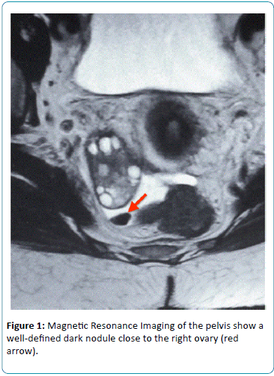
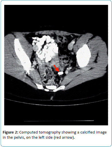
A transvaginal pelvic ultrasound was performed in December 2012 and the left ovary was not visualized. A pelvic MRI confirmed the absence of the left ovary, and a calcified structure with 1.1 cm posterior to the right ovary was identified, which was compatible with peritoneal loose body (Figure 1). Computerized tomography of the abdomen and pelvis confirmed the finding of the pelvic MRI. However, the calcified structure was at different location from the previous test (Figure 2), which reinforced the hypothesis of peritoneal loose body. The left ovary was not observed in the CT scan.
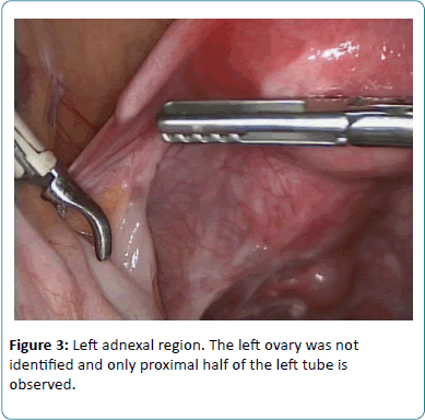
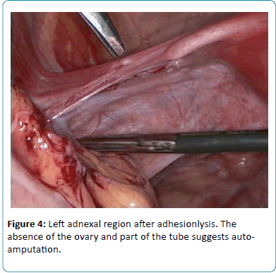
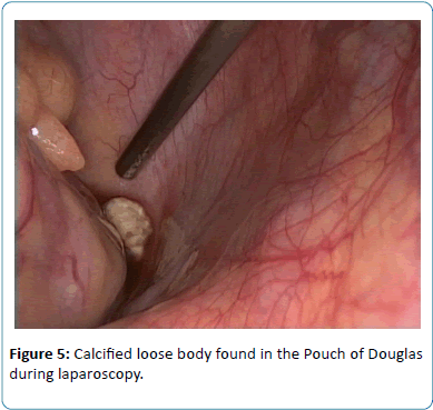
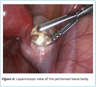
The patient underwent Laparoscopy to complete the investigation and treat the eventual causes of pelvic pain. Intraoperative inventory revealed normal right adnexa, left uterine tube without its fimbriae and distal part (Figures 3 and 4), absence of the left ovary, peritoneal adhesions in left adnexa region, and identification of a 1.0 cm calcified peritoneal loose body in the cul-de-sac region (Figures 5 and 6).
Adhesionlysis was performed and the calcified loose body was removed. The specimen was sent for pathological examination (Figures 4 and 5).
The patient recovered uneventfully after surgery and was discharged after 48 hours with no complaints. After six days she returned to office with no complaints of pelvic pain.
Due to intraoperative findings, we made the hypothesis of adnexal torsion followed by the left adnexa auto-amputation. During the post-operative appointment the patient was asked about previous episode of acute pelvic pain.
She then remembered of an episode of a very strong acute pelvic pain prior to the onset of the chronic pelvic pain. She reported that in that specific episode she was evaluated in the emergency room, but was discharged after all the tests resulted normal, even though she remained with strong pain.
Result of pathology
Fibrous connective tissue with dystrophic calcification and metaplastic ossification.
Discussion
Unilateral absence of tube and/or ovary is an extremely rare condition occurring in 1:11241 women. The cause is either congenital or acquired. The main cause in patients who acquire this condition is adnexal torsion, which may result in the ovary auto-amputation. This is usually due to and misdiagnosis of the torsion. Although most cases of adnexa autoamputation are asymptomatic, the history of acute or chronic pelvic pain may exist [4,5].
Epidemiological studies conducted in the United States show that ovarian torsion is the fifth leading cause of gynecologic surgical emergency. After the torsion the tube or ovary may undergo ischemia. Adnexal edema and congestion increases the effect of the initial ischemia, which may lead to necrosis. These cases usually manifest as an acute abdominal pain, with progressive worsening and little signs of peritoneal irritation. Because these findings are not specific, there may be a delay in the diagnosis and the onset of surgical intervention [6].
Kusaka and Mikuni published in 2007 a study with 20 cases of ovarian auto-amputation, showing higher prevalence on right adnexa (16/20). One explanation for this may be that the sigmoid protect the left ovary from torsion. However this predisposition is not a consensus in the literature (Table 1) [6].
| Author | Age (yo) | Size (cm) | Localization | Amputed side | Pre-operative diagnosis | Ovarian tissue | Histology | Previous pain |
| AikateriniPeitsidou, 2009 | 33 | 8 x 5 cm | Fornix | Right | No | Yes | Dermoid cyst | |
| Damian L.S. Eustace, 1992 |
24 35 |
- - |
- |
Right Right | - - |
- | - | |
| Kaori Koga, 2010 | 33 | 2 x 3 cm | Vesico-uterine space | Left | No | No | Calcification and cholesterol crystals | Yes - at 9 years old |
| JonhAmodio, 2010 | Neonate | 6 x6 cm | Right iliac fossa | Left | No | Yes | Hemorrhagic cyst, ovarian tissue with infarction and calcification | No |
| SrideviSankaran, 2009 | 28 | 8 x 8 cm | Fornix | Right | No | Yes | Benign fibroid | Yes, 2 years ago |
Table 1: List of peritoneal loose body Case Reports available on the literature originated from the adnexa.
In the case reported above, the patient reported an acute episode of pain in the left iliac fossa about three years before the surgical procedure. At that time, the adnexal torsion was not diagnosed, the initial pain improved spontaneously but the patient remained with a daily chronic pain.
After review of the imaging studies performed by the patient, it was clear those two years before laparoscopy she still had her left ovary visible by transvaginal ultrasound, but with reduced dimensions compared to the right ovary. It probably evolved to its auto-amputation during this time, since the MRI in 2012 showed no signs of the left ovary and the presence of a calcified loose body in the pelvis.
Peritoneal loose bodies are usually of little importance, since the great majority of these patients are asymptomatic. The finding of a loose body is often made at autopsy or during the performance of abdominal surgery for any other reason. In symptomatic cases, it can be manifested by pain, dyspepsia or compression of adjacent organs, leading to intestinal obstruction and urinary retention. Feature sizes range from 0.5 cm to 2.0 cm, and are classified by some authors as giants when they exceed 4.0 cm or 5.0 cm in diameter. Because it is a rare event, some differential diagnosis should be raised, such as: calcified uterine leiomyoma, foreign body granuloma, biliary calculi and ovarian teratomas [1,3].
Considering that the etiologies of theses lesions are predominantly benign, there is no specific treatment suggested for asymptomatic patients. Surgical resection is indicated in symptomatic cases or to elucidate tumors of unknown origin or with suspect characteristics. The recommended surgical approach for these cases is laparoscopy [6].
Based on her past history of acute pelvic pain and on imaging findings showing the presence of the left ovary in pelvic ultrasound and its absence after two years from the initial pelvic pain, we concluded that the patient experienced an episode of left adnexal torsion and due to the lack of diagnosis of ovarian torsion on that occasion it resulted in the auto-amputation of the left adnexa. These structures were deposited in Douglas' pouch and the ischemia due to the autoamputation promoted the progressive replacement of the ovarian tissue by fibrous connective tissue and calcium.
This case report illustrates a possible outcome of a misdiagnosis of ovarian torsion and reinforces the importance of a proper evaluation of acute pelvic pain, even when pelvic imaging don’t suggests adnexal torsion.
References
- Jang JT, Kang HJ, Yoon JY, Yoon SG (2012) Giant peritoneal loose body in the pelvic cavity. J Korean SocColoproctol 28: 108-110.
- Nozu T, Okumura T (2012) Peritoneal loose body. Intern Med 51: 2057.
- Kim HS, Sung JY, Park WS, Kim YW (2013) A giant peritoneal loose body. Korean J Pathol 47: 378-382.
- Peitsidou A, Peitsidis P, Goumalatsos N, Papaspyrou R, Mitropoulou G, et al. (2009) Diagnosis of an autoamputated ovary with dermoid cyst during a Cesarean section. FertilSteril 91.
- Sankaran S, Shahid A, Odejinmi F (2009) Autoamputation of the Fallopian Tube after Chronic Adnexal Torsion. J Minim Invasive Gynecol 16: 219-221.
- Lima GR, Girao MB, Baracat EC (2009) No Title. Manole.
- Kusaka M, Mikuni M (2007) Ectopic ovary: a case of autoamputated ovary with mature cystic teratoma into the cul-de-sac. J ObstetGynaecol Res 33: 368-370.
Open Access Journals
- Aquaculture & Veterinary Science
- Chemistry & Chemical Sciences
- Clinical Sciences
- Engineering
- General Science
- Genetics & Molecular Biology
- Health Care & Nursing
- Immunology & Microbiology
- Materials Science
- Mathematics & Physics
- Medical Sciences
- Neurology & Psychiatry
- Oncology & Cancer Science
- Pharmaceutical Sciences
