Disseminated Endometriosis and Leiomyomatosis Following Power Morcellation
Hariton E, Bortoletto P, Walsh BW, Devente JE and Anchan RM
Hariton E1*#, Bortoletto P1#, Walsh BW1, Devente JE2 and Anchan RM1
1Department of Obstetrics, Gynecology and Reproductive Biology, Harvard Medical School, USA
2Department of Obstetrics and Gynecology, East Carolina University, USA
- *Corresponding Author:
- Hariton E
Department of Obstetrics
Gynecology and Reproductive Biology
Harvard Medical School, USA
Tel: (617) 732-4285
E-mail: ranchan@bwh.harvard.edu
Received date: August 06, 2017; Accepted date: August 09, 2017; Published date: August 12, 2017
Citation: Hariton E, Bortoletto P, Anchan RM, Devente JE, Walsh BW (2017) Disseminated Endometriosis and Leiomyomatosis Following Power Morcellation. J Reproductive Endocrinol Infert Vol. 2:23. doi: 10.21767/2476-2008.100023
Copyright: © 2017 Hariton E, et al. This is an open-access article distributed under the terms of the Creative Commons Attribution License, which permits unrestricted use, distribution, and reproduction in any medium, provided the original author and source are credited.
Introduction
A 45-year-old, gravida two, para two, presented to the emergency department with acute right sided pelvic pain. A CT scan demonstrated an 11 cm left adnexal solid and cystic pelvic mass, concerning for ovarian torsion. Her past surgical history was notable for a laparoscopic supracervical hysterectomy four years ago for benign indications with uncontained power morcellation.
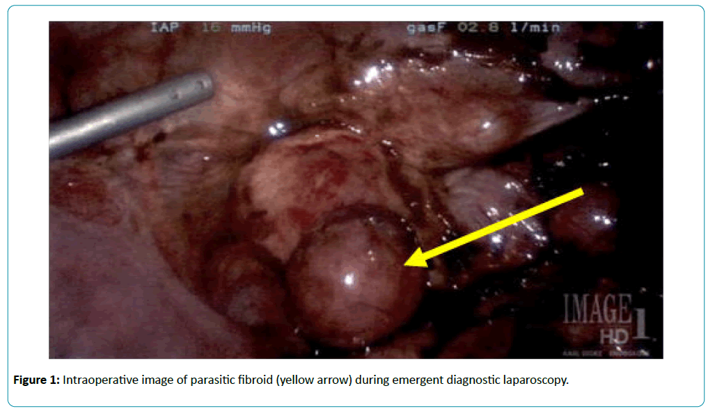
On exploratory laparoscopy, “chocolate fluid” was noted throughout the pelvis, concerning for recent rupture of an ovarian endometrioma. There were also approximately seven smooth, round, pink lesions present in the pelvis, which appeared to be parasitic leiomyoma leiomyomata, adherent to multiple structures (Figure 1). Histopathologic examination confirmed the presence of a hemorrhagic luteinized cyst and benign leiomyomas.
Surgeons considering a minimally invasive surgical approach without en bloc tissue removal should be aware of the potential complications associated with iatrogenic dissemination of benign viable endometrial and/or myometrial tissue associated with uncontained tissue morcellation [1-3].
References
- Cohen, A, Tulandi, T (2017) Long-Term sequelae of unconfined morcellation during laparoscopic gynecological surgery. Maturitas 97: 1-5.
- Meulen JFVD, Pijnenborg JM, Boomsma CM, Verberg MF, Geomini PM, et al. (2016) Parasitic myoma after laparoscopic morcellation: a systematic review of the literature. BJOG 123: 69–75.
- Heller DS, Cracchiolo B (2014) Peritoneal nodules after laparoscopic surgery with uterine morcellation: review of a rare complication. J Minim Invasive Gynecol 21:384-8.
Open Access Journals
- Aquaculture & Veterinary Science
- Chemistry & Chemical Sciences
- Clinical Sciences
- Engineering
- General Science
- Genetics & Molecular Biology
- Health Care & Nursing
- Immunology & Microbiology
- Materials Science
- Mathematics & Physics
- Medical Sciences
- Neurology & Psychiatry
- Oncology & Cancer Science
- Pharmaceutical Sciences

